


Intraocular Pressure: What It Is, What Is Normal, and Why It Matters
If your eye pressure was flagged at a recent appointment, you may be wondering what the number actually means and whether you need to be concerned. Intraocular pressure is one of the most important measurements in eye care, and understanding it can help you make sense of what you have been told and what comes next.
Quick Overview
- Intraocular pressure (IOP) is the fluid pressure inside your eye, and it plays a key role in protecting the optic nerve.
- Normal eye pressure falls between 10 and 21 millimetres of mercury (mmHg), though what is considered safe varies between individuals.
- Elevated eye pressure does not automatically mean glaucoma, but it is one of the most significant risk factors for developing it.
- Management ranges from prescription eye drops to laser treatment, depending on how high the pressure is and whether the optic nerve is already affected.
Below, we explain how eye pressure works, what raises it, and what can be done when it stays elevated.
What Is Intraocular Pressure?
 Your eye produces a clear fluid called aqueous humour. This fluid nourishes the lens, cornea, and other internal structures. It travels through each part of the eye that has no blood supply, then drains out through tiny channels at the front. The fluid inside the eye is constantly being made and drained. When those two processes stay in balance, pressure inside the eye stays stable.
When the drainage channels, known as the trabecular meshwork, become less efficient or blocked, fluid builds up faster than it can leave. Pressure rises. Untreated high eye pressure pushes against the optic nerve head at the back of the eye, and over time, that sustained pressure can cause the nerve damage associated with glaucoma.
What Is the Normal Range for Eye Pressure?
Normal eye pressure is generally between 10 and 21 millimetres of mercury (mmHg). Healthdirect Australia estimates that around 379,000 people in Australia are living with glaucoma, yet roughly half of those affected have never been diagnosed. Two large Australian population studies, the Blue Mountains Eye Study and the Melbourne Visual Impairment Project, identified that ocular hypertension affects roughly 3.7% of Australians aged over 40, meaning elevated eye pressure without any optic nerve damage yet.
That range is a guide, not a fixed rule. Some eyes handle higher pressures just fine. Others develop optic nerve damage even when pressure appears to be inside the normal range. That is why your ophthalmologist looks at pressure alongside the health of your optic nerve, your peripheral vision results, and your individual risk factors, not just a single number.
When IOP is considered elevated
Pressure consistently above 21 mmHg is no longer considered normal and is called ocular hypertension. People in this category are sometimes referred to as glaucoma suspects because they are more likely to develop glaucoma over time, though many never go on to develop it. The Ocular Hypertension Treatment Study found that early treatment with pressure-lowering eye drops cut the rate of patients developing glaucoma roughly in half over five years, from around 9.5% down to 4.4%.
Normal tension glaucoma
Here is something that surprises many people: not all glaucoma involves high eye pressure. Normal tension glaucoma is when the optic nerve is damaged even though IOP stays within the normal range. This can happen when the nerve is more fragile than usual, or when blood flow to the optic nerve is reduced. It is a good reminder that eye pressure alone does not tell the full story, which is why peripheral vision testing is part of a thorough assessment.
What Raises Intraocular Pressure?
A few different things can push eye pressure above the normal range.
Drainage obstruction
In primary open-angle glaucoma, the drainage angle is open, but the trabecular meshwork does not drain properly. Fluid builds up slowly, and pressure rises gradually. Most people have no idea this is happening. In angle closure, the drainage angle becomes physically blocked, pressure spikes rapidly, and treatment is needed urgently.
Blood pressure and vascular health
There is a relationship between blood pressure and eye health. The balance between blood pressure and intraocular pressure determines how much blood actually reaches the optic nerve. If blood pressure drops too low, the optic nerve may not receive enough supply even when IOP looks normal. Poorly controlled high blood pressure can affect fluid balance inside the eye over time.
Lifestyle factors
Some everyday habits have a modest effect on IOP. Reducing caffeine intake, maintaining a balanced diet, and staying physically active are all associated with small reductions in eye pressure for some people. Yoga poses that involve prolonged head-down positions, such as headstands, can temporarily raise IOP. These steps support overall eye health but are not a substitute for treatment when IOP remains elevated.
Family history and risk factors
If a close relative has glaucoma, your own risk of developing elevated IOP is higher. The risk of glaucoma increases further with age, short-sightedness, thin corneas, and conditions such as diabetes. If you have several risk factors, your ophthalmologist may recommend earlier or more frequent monitoring than standard guidelines suggest.
How Is Eye Pressure Measured?
 Eye pressure, or IOP, is measured using tonometry. There are several ways to measure eye pressure at a comprehensive eye examination. The most reliable method is contact tonometry, where a small instrument gently touches the surface of your numbed eye and measures the force needed to flatten a tiny area of the cornea. Non-contact tonometry, which most people know as the air puff test, works on the same principle without touching the eye.
A single reading does not give the complete picture. IOP measurements taken over time are far more informative than any one result. IOP can shift throughout the day, often sitting higher in the morning. Multiple readings taken at different times, combined with optic nerve imaging and visual field testing, give your ophthalmologist a far more accurate assessment of your eye health.
How Is Elevated Intraocular Pressure Managed?
The aim of treatment is to bring eye pressure down to a level where the optic nerve is protected. What that target looks like depends on the individual. Some people need moderate reductions; others need their pressure brought quite low to prevent further nerve damage and disease progression.
Prescription eye drops
For most people, prescription eye drops are the starting point. They work by either reducing fluid production inside the eye or improving how efficiently it drains through the trabecular meshwork. Drops are used daily, and the type is chosen based on how well they work for each person and how well they are tolerated.
Selective laser trabeculoplasty
Selective laser trabeculoplasty, or SLT, uses a laser to stimulate the trabecular meshwork and improve drainage, which helps lower eye pressure. It is typically considered when drops are not bringing IOP down enough, or when a patient finds it difficult to use drops consistently. SLT is done in two short sessions per eye, causes little to no discomfort, and works well for most glaucoma patients.
Surgical options
When drops and laser treatment are not enough, surgery may be recommended. A trabeculectomy creates a new drainage pathway so fluid can leave the eye more effectively. Surgery is generally reserved for advanced or poorly controlled glaucoma where other approaches have not achieved the required pressure reduction.
Getting Your Eye Pressure Assessed
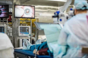 High eye pressure feels like nothing. There is no sensation, no blurry vision in most cases, and no warning sign you can notice yourself. That is what makes regular monitoring so important. Glaucoma caught early is far more manageable, and the earlier elevated pressure is identified, the more options are available to prevent vision loss.
At Armadale Eye Clinic in Melbourne, our team assesses IOP alongside optic nerve imaging and visual field testing to give you a complete picture of your eye health. To discuss your eye pressure readings or arrange an assessment, please call us on (03) 9070 5753.
Frequently Asked Questions
Can high eye pressure go away on its own?
A single elevated reading may reflect the time of day or recent caffeine intake rather than a persistent problem. Consistently elevated IOP, however, does not typically resolve without intervention. If your readings remain elevated across multiple visits, management is usually recommended to protect the optic nerve from progressive damage.
Is there a link between eye pressure and headaches?
A sudden severe spike in eye pressure, as can happen in angle closure glaucoma, can cause eye discomfort, blurry vision, and headaches that need urgent treatment. The gradual pressure elevation seen in primary open-angle glaucoma, on the other hand, typically causes no discomfort or headaches at all. If you experience sudden severe eye discomfort alongside a headache, seek urgent assessment.
Can lifestyle changes lower intraocular pressure meaningfully?
Reducing caffeine intake, staying active, and avoiding prolonged head-down positions may contribute to modest IOP reductions in some people. These habits support overall eye health and can complement clinical management, but they are not a substitute for prescribed treatment when IOP remains elevated.
How often should IOP be checked?
It depends on your individual risk. People with no risk factors and normal readings are typically reviewed every one to two years. Those with ocular hypertension, confirmed glaucoma, or other risk factors may need appointments every three to six months. Your ophthalmologist will recommend a schedule based on your findings.
Can glaucoma develop even after IOP is brought under control?
Yes, for some people. Lowering IOP significantly reduces the rate of glaucoma progression, but it does not eliminate the risk entirely. Some patients continue to show slow optic nerve changes even when pressure is at target, which is why ongoing monitoring of the nerve and visual field remains part of long-term care.
Note: Any surgical or invasive procedure carries risks. Before proceeding, you should seek a second opinion from an appropriately qualified health practitioner.
References
https://www.mdpi.com/2227-9059/12/10/2212
https://www.sciencedirect.com/science/article/pii/S2667376225000149

Multifocal intra-ocular lenses offer the opportunity to have cataract surgery and have great vision for distance, intermediate and near. There are a few golden rules Read more

We all develop presbyopia when we get to between 45-50 yeas of age. Presbyopia is the loss of the ability to focus up near. This Read more

The eye is an amazing organ with multiple parts all working together to receive light, convert it to neural signals, and send this signal onwards Read more

Unfortunately, no, long sightedness is not exactly as simple as being the opposite of short sightedness. People with long sightedness can in fact often still Read more

 Cataract vision refers to the gradual change in sight
 A comprehensive eye examination is a reliable way to detect cataracts. An eye specialist will assess your vision and examine the lens for signs of clouding.
 Understanding cataract symptoms, risk factors, and


